Diagnosis for Carcinoma
Breast:
- Mammogram
- Ultrasound Breast
- MRI
- PET scan
Combined mammography, clinical examination, and MRI
are more sensitive than any other individual test or combination
of tests.
| Modality |
Sensitivity |
Specificity |
Positive predictive value |
Indications |
Mammography
|
63-95%
(>95% palpable,
50% impalpable,
83-92% in women older than 50 y) (decreases to 35%
in dense breasts)
|
14-90%
(90% palpable)
|
10-50%
(94% palpable)
|
Initial investigation for symptomatic breast in women
older than 35 years and for screening; investigation of
choice for microcalcification
|
| Ultrasonography |
68-97% (palpable)
|
74-94% (palpable)
|
92% (palpable)
|
Initial investigation for palpable lesions in women
younger than 35 years
|
MRI
|
86-100%
|
21-97%
(< 40% primary cancer)
|
52%
|
Scarred breast, implants, multifocal lesions, and borderline
lesions for breast conservation; may be useful in screening
high-risk women
|
Scintigraphy
|
76-95% (palpable)
52-91% (impalpable) |
62-94%
(94% impalpable) |
70-83%
(83% palpable,
79% impalpable) |
Lesions larger than 1 cm and axilla assessment; may
help predict drug resistance
|
PET scanning
|
96%
(90% axillary metastases) |
100%
|
|
Axilla assessment, scarred breast, and multifocal lesions
|
Confirmation of Diagnosis:
- Fine Needle Aspiration Cytology (FNAC)
- Trucut or core biopsy
- Sterotactic biopsy under mammographic guidance
 Treatment
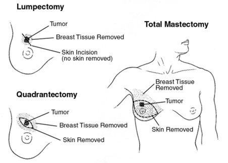
Surgery is considered primary treatment for breast cancer, as
many patients with early-stage disease are cured with surgery
alone. The goals of breast cancer surgery include complete resection
of the primary tumor with negative margins to reduce the risk
of local recurrences, and pathologic staging of the tumor and
axillary lymph nodes for providing necessary prognostic information.
Several different types of operations are available for the
treatment of breast cancer.
Adjuvant treatment for breast cancer involves radiation therapy
and a variety of chemotherapeutic and biologic agents.
Prognosis
Numerous prognostic and predictive factors for breast cancer
have been identified by the College of American Pathologists
(CAP) to guide the clinical management of women with breast
cancer.
Breast cancer prognostic factors include the following:
- Axillary lymph node status
- Tumor size
- Lymphatic/vascular invasion
- Patient age
- Histologic grade
- Histologic subtypes (eg, tubular, mucinous [colloid],
papillary)
- Response to neoadjuvant therapy
- ER/ PR status
- HER2 gene amplification and/or overexpression
Breast cancer predictive factors include the following:
- ER/PR status
- HER2 gene amplification and/or overexpression
Cancerous involvement of the lymph nodes in the axilla is an
indication of the likelihood that the breast cancer has spread
to other organs. Survival and recurrence are independent of
level of involvement but directly related to the number of involved
nodes.
Patients with node-negative disease have an overall
10-year survival rate of 70% and a 5-year recurrence
rate of 19%. In patients with lymph nodes that are positive
for cancer, the recurrence rates at 5 years are higher (30-70%)
Hormone-positive tumors have a more indolent course and are
responsive to hormone therapy.
Five-year survival rates are highly correlated with tumor stage,
as follows:
- Stage 0: 99-100%
- Stage I: 95-100%
- Stage II: 86%
- Stage III: 57%
- Stage IV: 20%
This prognostic information can guide physicians in making therapeutic
decisions. Pathologic review of the tumor tissue for
histological grade along with the determination of estrogen/progesterone
receptor status and HER2 status is necessary for determiningprognosis.
Evaluation of lymph node involvement by sentinel lymph node
biopsy or axillary lymph node dissection is generally necessary
as well. |